Heparan Sulfate Antibodies
10E4, 3G10, and JM403
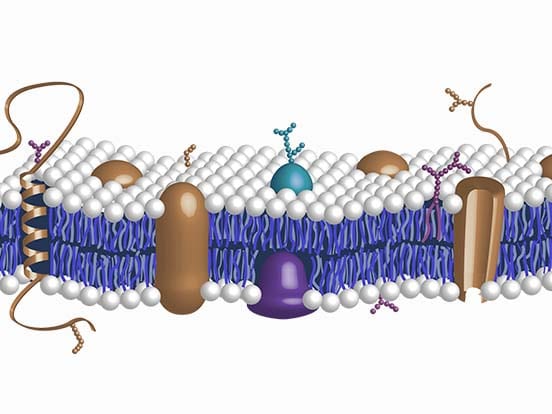
Heparan sulfate is a highly sulphated polysaccharide, synthesized as the glycosaminoglycan component of heparan sulfate proteoglycans (HSPGs). It is widely distributed on the cell surfaces and basement membranes in mammals. It is involved in important biological processes due to it displaying specific interactions with many biologically active proteins. This includes examining of the dynamic distribution of HSPG in tissues and analysing HS structures for the elucidation of HSPG biological functions.
What We Offer:
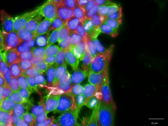
The non-immunogenic character of HS makes this type of antibody difficult to raise, so the few hybridoma-derived mouse anti-HS antibodies such as JM403, 10E4 and 3G10 are valuable tools for HS research. Using any of our multiple heparan sulfate antibodies that recognize subtle differences in HS patterning can be useful in applications such as flow cytometry allowing multiple cell types to be directly compared.
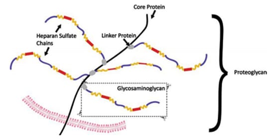
AMSBIO offers quality heparan sulfate antibodies from the important clones (JM403, 10E4 and 3G10), which are ideal for the binding of HS for HSPG research. AMSBIO also provides various anti-HS antibodies, recognizing distinct HS substructures and well characterized in previous studies. The 10E4 antibody is now available in biotinylated formats.
How 10E4, JM403, 3G10 antibodies and heparinase III enzymes work together
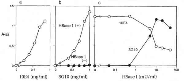
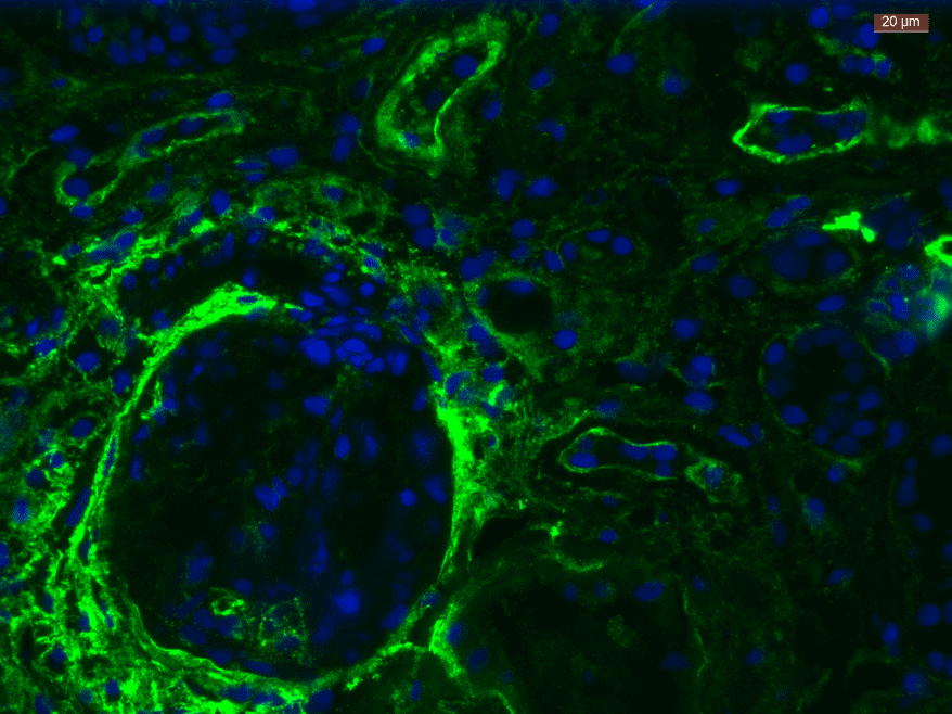
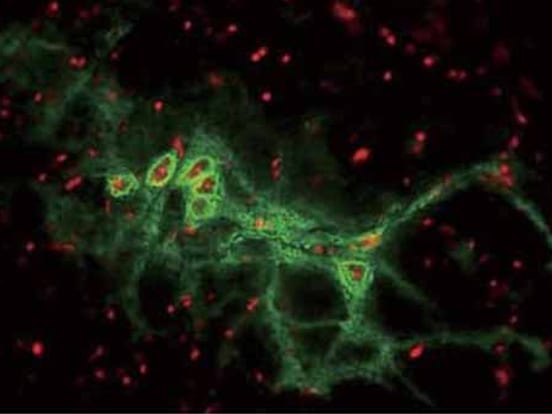
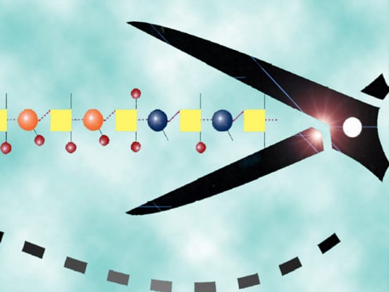
Clones 10E4 and JM403 recognize common epitopes on heparan sulfate (HS), which can be found on both basement membrane and cell-associated HS species. The 3G10 clone recognizes a neo-epitope exposed by digestion with heparinase III (HSase I) enzyme as seen in figure 3 (which destroys reactivity to 10E4 and JM403); so that the 3G10 clone can be used as control to 10E4 or JM403 (and vice versa).
10E4 Antibody
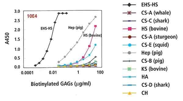
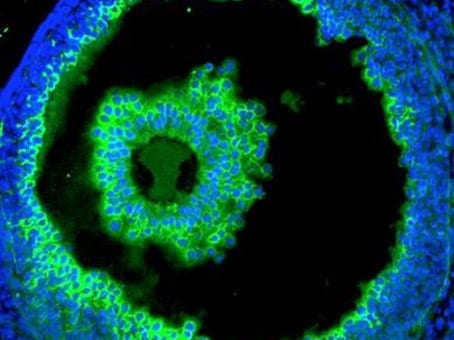
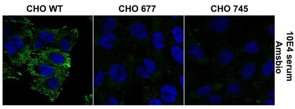
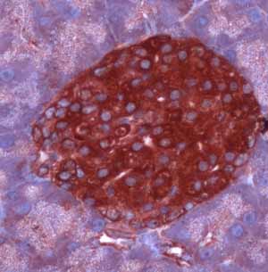
The monoclonal antibody 10E4 (F58-10E4 clone), which recognises the 10E4 epitope containing N-unsubstituted glucosamine, is commonly used to trace heparan sulfate proteoglycans. The 10E4 epitope also contains N-sulfated residues that are critical for the reactivity of the F58-10E4 antibody. The reactivity of heparan sulfate with this antibody is nearly nullified after the glycosaminoglycan has been treated with bacterial heparatinase I (Heparinase III) from Flavobacterium heparinum (EC 4.2.2.8). The 10E4 antibody does not react with hyaluronan, chondroitin sulfate, dermatan sulfate, keratan sulfate or DNA.
Examples of 10E4 Antibody Applications:
3G10 Antibody
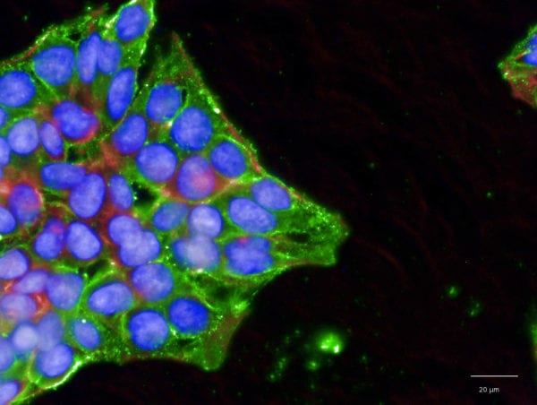
Anti-Heparan sulfate antibody (F69-3G10 clone) reacts with heparan sulfate neo-epitope 3G10, generated by digesting heparan sulfate with heparitinase I (Heparinase III) from Flavobacterium heparinum (EC 4.2.2.8). The desaturated hexuronate (glucuronate) located at the non-reducing end of the heparan sulfate fragments created by the enzyme is critical for the reactivity of the antibody. The 3G10 antibody does not react with intact heparan sulfate, oligosaccharides generated from chondroitin sulfates with bacterial chondroitinase ABC or AC, or generated from heparan sulfate with heparinase (Heparinase I): EC 4.2.2.7). There is only one 3G10 epitope on the HS stub which can only be recognized by one antibody at a time which allows these to be utilized as a tool for measuring how many chains are present per cell.
JM403 Antibody
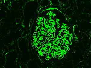
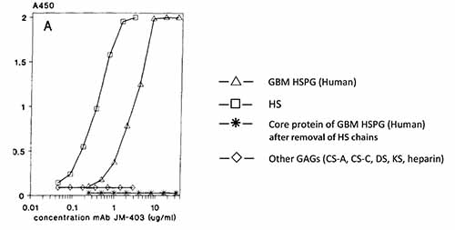
Heparan sulfate antibody (clone JM403) reacts with an epitope present in many types of heparan sulfate. The JM403 epitope includes (an) N-unsubstituted glucosamine residue(s) that are critical for the reactivity of the antibody. The reactivity of the JM403 antibody with most heparan sulfates is nearly completely abolished after treatment of the glycosaminoglycan with bacterial Heparitinase I (Heparinase III) from Flavobacterium heparinum (EC 4.2.2.8). The JM403 antibody does not react with heparin, hyaluronan, chondroitin sulfate, dermatan sulfate and keratan sulfate.
Citations
10E4 & 3G10 Antibodies
Differential expression of multiple cell-surface heparan sulfate proteoglycans during embryonic tooth development
Bai, X.M., et al (1994). Journal of Histochemistry & Cytochemistry, 42(8), 1043-1054.
Developmental changes in heparan sulfate expression: in situ detection with mAbs.
Bai, D.G., et al (1992). The Journal of Cell Biology, 119(4), 961-975.
Quantification of Malignant Breast Cancer Cell MDA-MB-231 Transmigration Across Brain and Lung Microvascular Endothelium.
Fan, J., & Fu, B.M. (2015). Annals of Biomedical Engineering, 1-13
Assessment of Proteins Associated With Complement Activation and Inflammation in Maculae of Human Donors Homozygous Risk at Chromosome 1 CFH-to-F13BCFH-to-F13B Locus and Complement Activation in Macula.
Keenan, T.D., et al (2015). Investigative Opthamology & Visual Science, 56(8), 4870-4879
Heparan sulfate and heparanase play key roles in mouse β cell survival and autoimmune diabetes.
Ziolkowsi, A.F., et al (2012). The Journal of Clinical Investigation, 122(1), 132.
Exocytosis of endothelial lysosome-related organelles hair-triggers a patchy loss of glycocalyx at the onset of sepsis.
Zullo, J.A., et al (2016). The American Journal of Pathology, 186(2), 248-258.
10E4, 3G10 and 3B3 Antibodies
The CS Sulphation Motifs 4C3, 7D4, 3B3 [-]; and Perlecan Identify Stem Cell Populations and Niches, Activated Progenitor Cells and Transitional Tissue Development in the Fetal Human Elbow.
Hayes, A.J., et al (2016) Stem Cells and Development, 25(11) 836-47.
10E4 & JM-403 Antibodies
Novel heparan sulfate structures revealed by monoclonal antibodies.
Van Den Born, J., et al. (2005) Journal of Biological Chemistry, 280(21), 20516-20523.
10E4, 3G10 & JM-403 Antibodies
Use of flow cytometry for characterization and fractionation of cell populations based on their expression of heparan sulfate epitopes.
Holley, R.J., et al. (2015) Glycosaminoglycans: Chemistry & Biology, 239-251.
Presence of N-unsubstituted glucosamine units in native heparan sulfate revealed by a monoclonal antibody.
Van Den Born, J., et al. (1995) Journal of Biological Chemistry, 270(52), 31303-31309.
Differential Expression of Proteoglycans by Corneal Stromal Cells in KeratoconusProteoglycan Expression in Keratoconus Corneal Stroma.
García, B., et al (2016) Investigative Ophthalmology & Visual Science, 57(6), 2618-2618.
JM-403 Antibody
Heparan Sulfate Structure: Methods to Study N-Sulfation and NDST Action.
Dagälv, A., et al (2015) Glycosaminoglycans: Chemistry & Biology, 239-251.
A monoclonal antibody against GBM heparan sulfate induces an acute selective proteinuria in rats.
Van Den Born, J., et al (1992) Kidney International, 41(1), 115-123
Monoclonal antibodies against the protein core and glycosaminoglycan side chain of glomerular basement membrane heparan sulfate proteoglycan: characterization and immunohistological application in human tissues.
Van Den Born, J., et al (1994) Journal of Histochemistry & Cytochemistry, 42(1), 89-102.
