This competition is now closed.
Do you use AMSBIO products? Then this competition is for you! Submit an original scientific image using any AMSBIO product and be in with a chance to win up to $500. Submit your image (or images) below. Winners will be chosen on the highest number of votes, so make sure you get your friends and colleagues to vote for you. You can also enter multiple images. Closing date is 31st October 2023. We’re really looking forwards to seeing them!
- 1st Prize = $500
- 2nd Prize = $250
- 3rd Prize = $100
- 3 x Runner's up prize of $50 each.
Ready to choose your favorites? You can vote for as many photos as you like, but only once per photo. Don't forget to come back later to see if there are any more photos you'd like to vote for!
Adele MucciMolecular BiologySwiss roll of a mouse colon with spontaneous Inflammatory Bowel Disease here ameliorated by a treatment with an innovative cell therapy approach. Stained with Hematoxylin Eosin to evaluate tissue damage and inflammation.
Adele MucciMolecular BiologySwiss roll of a mouse colon with spontaneous Inflammatory Bowel Disease here ameliorated by a treatment with an innovative cell therapy approach. Stained with Hematoxylin Eosin to evaluate tissue damage and inflammation.
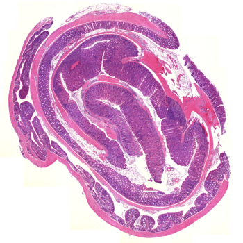
Adele Mucci Molecular BiologySwiss roll of a mouse colon with spontaneous Inflammatory Bowel Disease here ameliorated by a treatment with an innovative cell therapy approach. Stained with Hematoxylin Eosin to evaluate tissue damage and inflammation.

Matea BrezakCells & Cell CultureMammary gland epithelial organoid cultivated in 3D extracellular matrix after being frozen in Cellbanker2.
Matea BrezakCells & Cell CultureMammary gland epithelial organoid cultivated in 3D extracellular matrix after being frozen in Cellbanker2.
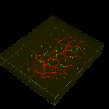
Matea Brezak Cells & Cell CultureMammary gland epithelial organoid cultivated in 3D extracellular matrix after being frozen in Cellbanker2.

Gabriele CasiratiProteins & PeptidesCytokines
Gabriele CasiratiProteins & PeptidesCytokines
Gabriele Casirati Proteins & PeptidesCytokines
Michael RehmanCells & Cell CulturePrimary cilia (green) ZO1 (red) in renal primary cells. Primary cells frozen and thawed with Cellbanker.
Michael RehmanCells & Cell CulturePrimary cilia (green) ZO1 (red) in renal primary cells. Primary cells frozen and thawed with Cellbanker.
Michael Rehman Cells & Cell CulturePrimary cilia (green) ZO1 (red) in renal primary cells. Primary cells frozen and thawed with Cellbanker.

Laurent GautronAntibodiesTomato-positive enteric neuron (red) in the mouse gut labeled with RFP antibody (cat#AB1140-100). Image acquired with confocal microscopy.
Laurent GautronAntibodiesTomato-positive enteric neuron (red) in the mouse gut labeled with RFP antibody (cat#AB1140-100). Image acquired with confocal microscopy.
Laurent Gautron AntibodiesTomato-positive enteric neuron (red) in the mouse gut labeled with RFP antibody (cat#AB1140-100). Image acquired with confocal microscopy.
Guilherme NaderCells & Cell CultureCancer cells often experience mechanical stress while moving or growing in solid tumors and such mechanical strains have been shown to impact the integrity of their nuclei. Aiming to recapitulate in vivo mechanical stress, Hap1 cells (human hepatic adenocarcinoma cell line) were confined to a height of 2µm (using microfabricated devices) and imaged using a Zeiss Confocal Airy Scan microscope under 63x magnification. Note nuclear envelope blebs and DNA herniations caused by the confinement. This phenotype has also been reported in patient samples of solid tumors. -Green: IRE1-GFP (a transmembrane ER protein) -Red: ER luminal marker with KDEL sequence at the C terminus fused to mCherry -Blue: Hoechst (DNA)
Guilherme NaderCells & Cell CultureCancer cells often experience mechanical stress while moving or growing in solid tumors and such mechanical strains have been shown to impact the integrity of their nuclei. Aiming to recapitulate in vivo mechanical stress, Hap1 cells (human hepatic adenocarcinoma cell line) were confined to a height of 2µm (using microfabricated devices) and imaged using a Zeiss Confocal Airy Scan microscope under 63x magnification. Note nuclear envelope blebs and DNA herniations caused by the confinement. This phenotype has also been reported in patient samples of solid tumors. -Green: IRE1-GFP (a transmembrane ER protein) -Red: ER luminal marker with KDEL sequence at the C terminus fused to mCherry -Blue: Hoechst (DNA)
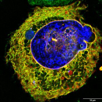
Guilherme Nader Cells & Cell CultureCancer cells often experience mechanical stress while moving or growing in solid tumors and such mechanical strains have been shown to impact the integrity of their nuclei. Aiming to recapitulate in vivo mechanical stress, Hap1 cells (human hepatic adenocarcinoma cell line) were confined to a height of 2µm (using microfabricated devices) and imaged using a Zeiss Confocal Airy Scan microscope under 63x magnification. Note nuclear envelope blebs and DNA herniations caused by the confinement. This phenotype has also been reported in patient samples of solid tumors. -Green: IRE1-GFP (a transmembrane ER protein) -Red: ER luminal marker with KDEL sequence at the C terminus fused to mCherry -Blue: Hoechst (DNA)

Jelte van der VaartCells & Cell CultureDifferentiating airway organoid thawed from CellBankerI
Jelte van der VaartCells & Cell CultureDifferentiating airway organoid thawed from CellBankerI
Jelte van der Vaart Cells & Cell CultureDifferentiating airway organoid thawed from CellBankerI