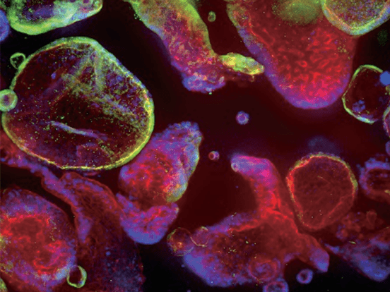
CollaFibr™ scaffold (green) with MEF DR4 cells, stained with Hoechst and phalloidin
CollaFibR™ Scaffold for Cell Culture
Dry Spun type I collagen fibers for 3D cell culture
3D cell cultures offer a more accurate representation of in-vivo conditions when studying cell and tissue models. Harnessing the power of dry-spinning technology, the CollaFibR™ scaffold consists of highly uniform and regulatory friendly collagen fibers. These matrices closely mimic the biomechanical and biochemical characteristics of natural collagen scaffolds, providing researchers with a more comprehensive understanding of cellular biology.
Features
- Produced using GMP bovine type I collagen, and resemble natural collagen fiber structures
- Degradable with collagenase for minimally invasive cell extraction/recovery
- User friendly 12-well plate inserts
- Compatible with brightfield, epifluorescence, confocal and live cell microscopy
- UV sterilized and ready to use on receipt
- Available with fluorescent tag
- Applications: Cellular Co-Cultures, 3D cytotoxicity studies, Cell Adhesion assays, Cell Migration assays, ECM deposition assays, Stem Cell Research and In-vitro models for High Throughput Screening

Product Information Table
| Name | Datasheet | Packsize | Order |
|---|---|---|---|
| CollaFibR Scaffold: No FITC/ with coverslip (Cross-Hatched Fibers) | Each (Individually Packaged Ring Insert, to fit 12 well plate) | View | |
| CollaFibR Scaffold: No FITC/ no coverslip (Cross-Hatched Fibers) | 12x (Individually Packaged Ring Inserts, to fit 12 well plate) | View | |
| CollaFibR Scaffold: No FITC/ no coverslip (Cross-Hatched Fibers) | Each (Individually Packaged Ring Insert, to fit 12 well plate) | View | |
| CollaFibR Scaffold: No FITC/ with coverslip (Cross-Hatched Fibers) | 12x (Individually Packaged Ring Inserts, to fit 12 well plate) | View | |
| Fluorescent CollaFibR Scaffold: FITC tag/ with coverslip (Cross-hatched Fibers) | 12x (Individually Packaged Ring Inserts, to fit 12 well plate) | View | |
| Fluorescent CollaFibR Scaffold: FITC tag/ with coverslip (Cross-hatched Fibers) | Each (Individually Packaged Ring Insert, to fit 12 well plate) | View |
3D Cell Culture on the CollaFibR™ Scaffold

Fluorescent-Advanced CollaFibR™ Scaffold for 3D Cell Culture has been seeded with fibroblasts (MEF DR4) which have infiltrated the collagen fiber scaffold. The cells are attached to the scaffold showing elongation and alignment with the collagen fibers. Imaged using epifluorescence microscopy, where the cells are stained with Hoechst and phalloidin.
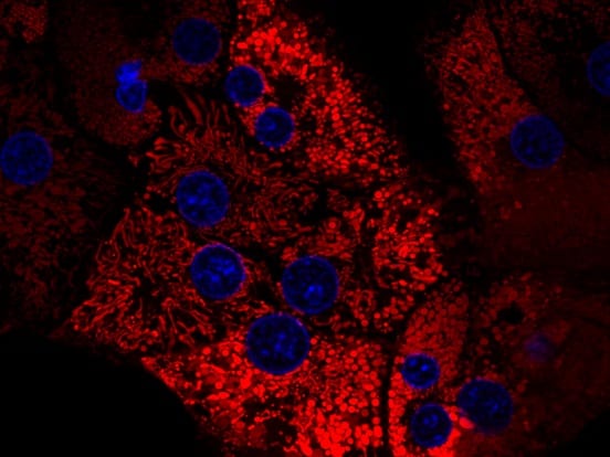
Basic-Advanced CollaFibR™ Scaffold for 3D Cell Culture is easily used in live cell confocal imaging. Tdtomato expressing primary tenocytes are seeded into the scaffold, which is stained using anti-collagen antibodies.
Versatile Imaging Capabilities
In CollaFibR™ Scaffolds, the collagen fibers drive consistent and elongated cellular morphologies, which are easily imaged using brightfield and epifluorescence microscopy. Matrigel poses limitations for staining and imaging analysis due to background interference from the coverslip and cells growing on the plate.

High Cell Viability on CollaFibR™ Scaffold

A wide range of primary cells, stem cells, and cell lines have been cultured on CollaFibR™, and all have over 90% cellular viability.

Aligned myotubes cultured on parallel CollaFibR™ scaffold
Try out the parallel CollaFibR™ scaffold with our Skeletal Muscle Differentiation kit. The kit uses a simple 3 step process of media change to differentiate human pluripotent stem cells to skeletal muscle with high yields and without cell sorting or genetic manipulation.
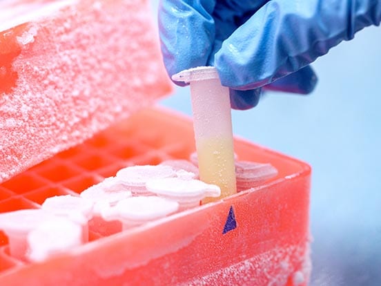
Collagenase Degradation of CollaFibR™ for Cell Recovery
Our Collagenase NB and Neutral Protease NB Enzymes are the perfect choice for degrading the CollaFibR™ scaffold for easy cell extraction.
- Recommended is Collagenase NB 4 Standard Grade or Collagenase NB1 Premium Grade
- Dependable batch-to-batch consistency
- Isolate high yields of viable cells
- Available in research grade, clinical grade, and animal-free options
CollaFibR™ Scaffold Frequently Asked Questions
What is the form-factor of CollaFibR™ scaffold?
CollaFibR™ scaffolds are supplied as individually packaged ring inserts which fit inside the wells of a standard 12-well cell culture plate.
What is the surface area of a CollaFibR™ scaffold and how cell culture media should be used?
The imageable surface area is 0.79 cm². Each insert holds up to 500μL of cell culture media.
How should CollaFibR™ scaffolds be stored?
CollaFibR™ scaffolds can be stored at room temperature (20-25°C) for up to 12 months in their original packaging.
Which CollaFibR™ scaffolds format should be used for microscopy?
The "basic" CollaFibR™ scaffold is non-fluorescent and housed in a clear medical grade polystyrene for standard optical microscopy. The "advanced" format can be bought with or without a fluorescent tag and has a glass bottom for confocal microscopy and other forms of light microscopy.
What fluorescent tags are available on CollaFibR™ scaffold?
Currently the fibers can be bought with a FITC tag.
Can cells be recovered from the CollaFibR™ scaffold?
Collagenease IV enzymatic degradation of the CollaFibR™ scaffold will release cultured cells for downstream applications. Please see (Collagenase degradation protocol) for recommendations.
Are the CollaFibR™ scaffolds sterile?
CollaFibR™ scaffold inserts are pre-sterilised and single packaged for ease of use. Re-sterilisation is not recommended.
What analytical modalities are compatible with CollaFibR™ scaffolds?
CollaFibR™ scaffold is compatible with live cell imaging, immunofluorescence for regular and high-resolution microscopy and minimally invasive cell recovery using Collagenase IV enzymatic degradation of the scaffold.
What is the source of collagen used in CollaFibR™ scaffolds?
Type I Collagen obtained from Bovine hide (skin) manufactured in a GMP facility.