Lentivirus for Nano-Lantern Luminescence Imaging
Enhanced bioluminescence imaging for real time cellular studies
Our range of orange nano-lantern lentivirus products has been developed using a proprietary lentiviral vector system and is ideal for in vivo luminescence Imaging. This range of ready-to-use lentivirus overcomes the limitations of fluorescent markers and the low brightness of traditional bioluminescence imaging to provide enhanced signal intensity bioluminescence imaging for real-time in vivo cellular observations. Perfect for intricate cellular research, high-throughput drug screening, and single-cell tracking in living animals and plants
Features
- Enhanced Bioluminescence: Nano-Lantern uses bioluminescence resonance energy transfer (BRET) to produce stronger signals than traditional bioluminescence imaging methods.
- No External Excitation: It doesn't require laser light activation, avoiding autofluorescence, phototoxicity, and photobleaching issues.
- Sub-Cellular Imaging: It provides bright and high-resolution imaging of small, rapidly moving cellular structures.
- Low Background Signal: Nano-Lantern offers high sensitivity by reducing background noise, enabling the detection of single molecules.
- Real-Time In Vivo Imaging: It allows dynamic visualisation of specific biological activities in living organisms.
Why use Nano-Lantern?
Fluorescent markers and bioluminescence are two widely utilized techniques for in vivo cell imaging. Fluorescence imaging is an important tool but relies on external laser light, which can introduce autofluorescence, phototoxicity, and photobleaching. Bioluminescence imaging does not require external light excitation but suffers from low brightness and requires longer exposure times, making it difficult to observe fast-moving cellular structures.
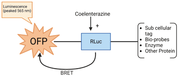
To address these limitations, an enhanced bioluminescence imaging method called Nano-Lantern was developed. This technique uses bioluminescence resonance energy transfer (BRET) to enhance the signal intensity of bioluminescence imaging. By introducing the substrate coelenterazine, energy transfer occurs between the fluorescent protein (FP) and luciferase, resulting in brighter bioluminescence signals. Refer to the scheme below for a visual representation of this process.
Nano-Lantern is bright enough to image sub-cellular structures and can even detect single molecules. It has an extremely low-level background signal, making it more sensitive and quantitative. When linked to a targeting protein, Nano-Lantern emits light in response to specific biological activity, making it a great tool for visualizing specific biological activity In vivo in real-time.
Nano-Lantern Bio-Probes
We have utilized proprietary lentiviral vector systems to generate a set of Orange Nano-Lantern Lentivirus products for In Vivo luminescence imaging applications. These Lentivirus products connect a well-defined organelle targeting structure or a Bio-probe to the specially designed Orange-Nano-Lantern structure. This Nano-Lantern consists of an enhanced Renilla-Luciferase connected to an Orange Fluorescent Protein (OFP). see the Lentivector’s Scheme below:
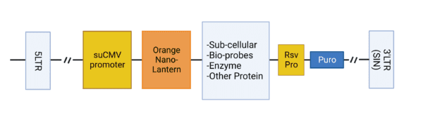
The specially designed Nano-Lanterns emit light in response to specific biological activities (such as Ca++ dynamics) or for In Vivo luminescence imaging of sub-cellular structures.
Orange Nano-Lantern Lentivirus products
| Name | Packsize | Order |
|---|---|---|
| Nano-Lantern Endoplasmic Reticulum (ER) | 1x10^8 IFU/ml x 200ul | View |
| Nano-Lantern Endosomes | 1x10^8 IFU/ml x 200ul | View |
| Nano-Lantern Golgi imaging | 1x10^8 IFU/ml x 200ul | View |
| Nano-Lantern Microtubule | 1x10^8 IFU/ml x 200ul | View |
| Nano-Lantern Nuclear-Membrane | 1x10^8 IFU/ml x 200ul | View |
| Nano-Lantern plasma membrane | 1x10^8 IFU/ml x 200ul | View |
| Nano-Lantern Cytoplasm area | 1x10^8 IFU/ml x 200ul | View |
| Nano-Lantern Cytoplasmic NAD+ Bioprobe | 1x10^8 IFU/ml x 200ul | View |
| Nano-Lantern H2B | 1x10^8 IFU/ml x 200ul | View |
| Nano-Lantern Lysosomes | 1x10^8 IFU/ml x 200ul | View |
| Nano-Lantern Mitochondria | 1x10^8 IFU/ml x 200ul | View |
| Nano-Lantern Mitochondrial NAD+ Bioprobe | 1x10^8 IFU/ml x 200ul | View |
| Nano-Lantern Nuclear Ca++ Bioprobe | 1x10^8 IFU/ml x 200ul | View |
| Nano-Lantern Nuclear Cas9 Probe | 1x10^8 IFU/ml x 200ul | View |
| Nano-Lantern Nuclear NAD+ Bioprobe | 1x10^8 IFU/ml x 200ul | View |
| Nano-Lantern Nuclear dCas9 Probe | 1x10^8 IFU/ml x 200ul | View |
| Nano-Lantern Nucleus area | 1x10^8 IFU/ml x 200ul | View |
| Nano-Lantern Zyxin | 1x10^8 IFU/ml x 200ul | View |
| Nano-Lantern beta-tubulin | 1x10^8 IFU/ml x 200ul | View |
| Nano-Lantern hTERT | 1x10^8 IFU/ml x 200ul | View |