Oris™ Pro Cell Migration Assays
The ideal cell migration assay for high-throughput screening applications
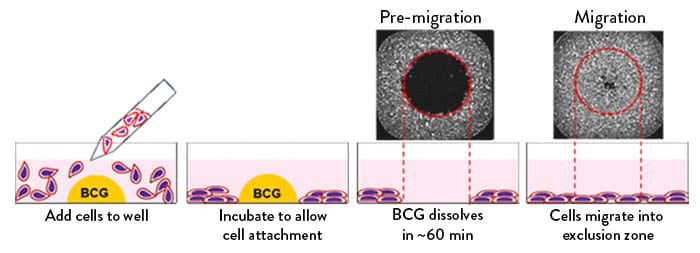
Oris™ Pro Assays use a water-soluble biocompatible gel (BCG) rather than a physical silicon barrier as in the Oris™ Assays, to create a cell-free detection zone for cell migration experiments. Cells are seeded around a spot of non-toxic BCG that dissolves after media and cells are added to each well, allowing for the rapid and easy quantification of cell migration.
Features
- Compatible with any adherent cell line
- Available with tissue culture treated plates or collagen I coated surfaces
- Compatible with liquid handlers, plate washers, high-content screening readers, and other high-throughput screening tools
Benefits
- Allows you to rapidly quantify cell migration/invasion in real time
- Significantly reduces handling due to the automation friendly design
- Creative design- use pre-coated assays or apply your own extracellular matrices
- Monitor phosphorylation events- perform in-cell westerns during cell migration
- Simplest assay set up and full automation
How does the Oris™ Pro Cell Migration Assay work?
In each well, a water-soluble Bio-Compatible Gel (BCG) creates a central cell-free Detection Zone for cell migration experiments. Upon attachment of the cells the BCG dissolves within 60 minutes, allowing the cells to migrate into the Detection Zone at the center of each well.
96-well plates
| Name | Packsize | Order | |
|---|---|---|---|
| We couldn't find any records. | |||
384-well plates
| Name | Packsize | Order | |
|---|---|---|---|
| We couldn't find any records. | |||
Frequently Asked Questions
What types of experiments can be performed with the Oris™ and Oris™ Pro Assays?
The Oris™ Cell Migration Assays can be utilized to detect chemokinesis, perform morphological analysis, and mimic a 2-D closure/wound assay. The invasion assays are designed to enable cell movement through an extracellular matrix in 3-Dimensions. The Oris™ Assays, however, are not suitable for chemotaxis or for use with non-adherent cell lines.
How do I know what ECM coating is best for my cell line?
Oris™ assays are available as TC treated, collagen I and fibronectin coated plate options for the Oris™ Stopper-Based Assays. Our TriCoated kit has 32 wells of each of these surfaces and is a good first option to study migration behavior of your cell lines on different ECMs. We also offer the Oris™ Universal Cell Migration Assembly Kit where you have the ability to apply your own custom ECM coating to the plate in your lab.
Oris™ Pro Assay kits are offered in tissue culture treated and collagen I coated formats.
Should the Oris™ Cell Seeding Stoppers be removed in order to add a substrate to the bottom of the plates and then reinserted?
An Oris™ Universal Cell Migration Assembly Kit is better suited for this purpose as it provides you with an empty plate available for coating and a separate packet of sterile stoppers that can be manually inserted after you apply an ECM of your choice.
Which assay format is better for me, Oris™ or Oris™ Pro?
The use of a Biocompatible Gel in Oris™ Pro assays instead of cell seeding stoppers permits complete access to the assay wells by automated liquid handling equipment at all steps of the assay from start to finish. No manual steps are necessary to add cells to the wells or to remove stoppers. The ability to run completely automated cell migration assays offers the benefits of both cost and time savings. Additionally, the high optical quality microplate provided with the Oris™ Pro assays is specifically designed for High Content Imaging.
The Oris™ Stopper-Based Assays are provided with a mask that make them uniquely well suited for use with microplate readers. The cell seeding stopper enables experiments where it is desirable for the cells to incubate in the plate with a reagent prior to initiating the experiment.
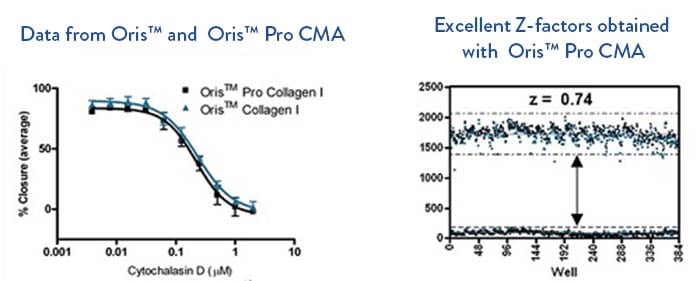
Platypus has tested multiple cell lines (HT-1080, MDA-MB-231 and HUVEC) with 4 different classes of migration inhibitors in parallel using Oris™ and Oris™ Pro assays and found no differences in migration or response to inhibitors.
How do I determine how many cells to seed in each well?
The goal is to achieve 95-100% confluence of the monolayers around the cell exclusion zone upon initiation of migration. Seeding at too high of a density is likely to result in cells in the Detection Zone at the beginning of the assay while seeding too few cells might result in delayed migration as the cells will need to fill in before migrating. It is best to test several different cell seeding densities when beginning to optimize the assay in order to find the best one as described in Appendix 1 in the assay protocol.
How long should I allow the cells to migrate or invade?
An advantage of the Oris™ Cell Migration and Invasion Assays is that the assay wells may be visually inspected at any time during the incubation period to ascertain how much the cells have moved into the Detection Zone. The first time you run an Oris™ assay, we recommend you periodically observe the assay wells under a microscope to assess the progression of migration/invasion into the Detection Zones. The ability of cells to migrate and their rate of migration depends upon the characteristics of a given cell type and the extracellular matrix (ECM) on which they are seeded. In order to achieve robust Z factors (>0.4), an endpoint time should be selected where the cells have closed at least 2/3 of the original open area of the Detection Zone. However, we suggest that the experiment be terminated prior to full closure of the Detection Zone.
The invasive phenotype of cells is cell line dependent. Invasion into the ECM overlay is usually observed within 24-72 hr. For extended experiments, we recommend that you change media above the ECM overlay and apply fresh inhibitors every 48-72 hr.
Is it possible to use only a partial assay plate and use the other wells for another experiment?
The Oris™ Cell Seeding Stoppers are grouped into strips comprised of 4 stopper tips. This allows the user to remove any factor of 4 stoppers at a time and leave the remainder of the stoppers available for future experiments. You should cover unused wells with a sterile plate sealer to protect from contamination. Limit the number of partial experiments you perform with plates that are ECM coated as temperature extremes may alter integrity of protein coatings. Care should also be taken when using fixatives as these volatile chemicals may react with adjacent unused assay wells. Store the partially used plates back in the original vapor barrier pouch at 4°C.
The flexibility to use partial plates depends on the assay. The Oris™ Cell Migration Assembly Kit-FLEX offers the most convenient option for partial plate experiments with the ability to perform up to 4 separate 24 well experiments with the materials provided.
For Oris™ Pro, we recommend that you do not run partial plate experiments as humidity in the incubator may affect BCG dissolution in the unused wells.
Are the Oris™ Cell Seeding Stoppers reusable with new sterile plates?
We do not recommend reusing Oris™ Cell Seeding Stoppers as sterilization procedures may alter stopper sealing properties or the stoppers may absorb and leach the chemicals used for sterilization. The Oris™ Cell Seeding Stoppers cannot be re-autoclaved as the heat and pressure will deform the stoppers and cause them to become brittle.
The Oris™ Cell Seeding Stoppers are specifically designed for the geometry of the 96-well plate that is provided to you in the Oris™ assay kits. The proper fit and seal will not be achieved in other 96-well microplates.
How can I tell the difference between cell migration and proliferation in the Oris™ Assays?
Generally speaking, cell proliferation is a factor of a given cell's doubling time. One step that you can take to ensure that your cells are not doubling in the assay is to run the assay for less time than the cells require for doubling. Another way to determine if proliferation is occurring during the migration period is to immunostain with an anti- Ki67 antibody, a marker only found on proliferating cells.
How do I test siRNA knock down cells in the assay?
There are two approaches that you might take. First, you could transfect the cells separately then transfer to either the Oris™ Stopper-Based Assay or the Oris™ Pro Assay. Alternatively, you could transfect your cells directly the in Oris™ assay plate with the stoppers still in place as described by Jiang, et al., Int. J. Cancer: 127, 505-512 (2010).
At what point can I add test compounds to be evaluated in the assays?
Test compounds may be added to the media once the cells have attached and spread following removal of the stoppers in the Oris™ Stopper-Based Assays. For Oris™ Pro Assays, test compounds may be added once the BCG has dissolved and the cells have fully attached and spread, but before migration takes place. This adhesion time is cell line and ECM dependent with attachment times ranging from 1 hr on Collagen I coated surfaces to 3 hr on TC treated surfaces. You may wish to remove and replace the medium prior to adding test compounds in order to eliminate any non-adherent cells. In the invasion assays, compounds may be incorporated into the BME or collagen I overlay solutions or added to the medium that is added above the overlay.
What stains and dyes should I use to assess migrating/invading cells in the Oris™ Assays?
Near-IR and fluorescent dyes derived from the fluorescein, rhodamine and cyanine families can all be used with this system as long as your detection system has the appropriate excitation and emission filters. The Oris™ Stopper-Based and Oris™ Pro Assays do not restrict your flexibility in staining cells. You can use multiple stains as long as the emission wavelengths of the dyes do not overlap. This capability of supporting multiplexed analysis is an attractive attribute of the Oris™ assays.
A nuclear stain such as DAPI offers the best choice for accurately enumerating cells within an Oris™ Assay Detection Zone. Nuclei appear as distinct, punctate dots which can be easily captured using ImageJ or High Content Imaging software. This approach, however, may not work with microplate readers due to its lower intensity and lack of instrument sensitivity. Other stains such as TRITC-phalloidin and Calcein AM may be used to stain the F-actin filaments and cytoplasm, respectively, and can be used in area closure assessments.
What options are there for prelabeling cells prior to seeding in the Oris™ Assays?
Cells may be prelabeled with dyes such as CellTracker Green™ or DiI (Life Technologies) in accordance with the manufacturer's protocols prior to seeding in Oris™ Assays. It is prudent to ascertain that the dyes do not affect cell motility by running parallel assay wells using unlabeled cells. Alternatively, cell lines may be used that stably express fluorescent proteins, such as GFP.
What are the best ways to establish pre-migration or pre-invasion reference wells in the Oris™ Stopper-Based Assays?
The Oris™ Cell Seeding Stoppers act as physical barriers that prevent the cells from moving into the Detection Zones until they are removed from assay wells. You can designate several pre-migration reference wells in which the stoppers will remain in place until results are read. For longer assays, you may need to seed the designated reference wells at a lower density so that the cells in the reference wells do not overgrow prior to the end of the experiment. Using this approach, it is necessary to seed cells in test and reference wells at different concentrations. Seed test wells at the optimal density to achieve 95-100% confluence upon stopper removal, but seed reference wells at a sub-optimal density to achieve 50-75% confluency. These reference wells will remain populated with Oris™ Cell Seeding Stoppers until the end of the assay whereas stoppers will be removed from test wells that will receive an overlay of ECM to initiate the invasion assay. After the appropriate incubation time to allow for invasion in the test wells, the Oris™ Cell Seeding Stoppers are then removed from the reference wells, the wells overlaid with ECM and allowed to gel. At this point, the entire plate containing both the test and reference wells can be stained and analyzed.
As Oris™ Pro Assays do not have stoppers that can be left in place during the assay, how can I establish pre-migration or pre-invasion references?
There are several appropriate options for establishing pre-migration/pre-invasion references in the Oris™ Pro assays: 1) A separate assay plate or assay wells can be fixed after the BCG has dissolved and the cells have attached and spread. These wells can then be stained with the rest of the wells once migration/invasion has taken place. 2) Pre-migration/pre-invasion images may be captured of all assay wells after the BCG has dissolved and the cells have attached and spread. This may be done in conjunction with cells labeled with CellTracker Green™ or expressing GFP. 3) Cells in several assay wells may be treated with a concentration of Cytochalasin D, an actin polymerization inhibitor, which completely arrests cellular motility and thus serves to 'freeze' the cells around the Detection Zone. Investigators might want to try several concentrations of Cytochalasin D ranging from 0.5uM- 2uM to select one that is not cytotoxic to their particular cell line.
What is the purpose of designating pre-migration/pre-invasion reference wells within Oris™ and Oris™ Pro Assay plates?
Pre-migration and pre-invasion references wells are used in order to establish the size of the Detection Zone before cells have moved in. For stopper-based assays, it is easy to establish at least 4 wells where the stoppers remain in place on each plate until the end of the assay because stoppers are provided in strips of 4.
For Oris™ Pro assays, pre-migration and pre-invasion references can be established in any number of assay wells by using an inhibitor such as Cytochalasin D at a concentration that completely arrests cell movement. Pre-migration and pre-invasion references can also be established by capturing images of each well after the BCG is dissolved for direct comparison at the end of the assay. It is prudent to establish several separate pre-migration or pre-invasion reference wells for each cell type if you are testing multiple cell lines on a single Oris™ Pro Assay plate.
What are the plate dimensions for Oris™ and Oris™ Pro plates?
| Oris™ Plates (96-well w/ Stoppers) | Oris™ Pro Plates (96-well w/ BCG) | Oris™ Pro 384 Plates (384-well w/ BCG) | |
|---|---|---|---|
| Diameter of well- bottom (mm) | 6.30 | 6.58 | 3.30 |
| Diameter of well- top (mm) | 6.45 | 6.96 | 3.70 |
| Plate height (mm) | 14.90 | 14.40 | 14.4 |
| Plate height with lid (mm) | 23.90 | 17.00 | - |
| Offset of wells (A-1 location, X) (mm) | 14.40 | 14.38 | 12.13 |
| Offset of wells (A-1 location, Y) (mm) | 11.20 | 11.24 | 8.99 |
| Distance between wells (mm) | 9.00 | 9.00 | 4.50 |
| Well depth (mm) | 12.20 | 10.90 | 11.50 |
| Thickness of well bottom (µm) | 250 | 190 +/-10 | 190 +/-10 |
| Suggested cell seeding volume per well (µL) | 400 | 392 | 138 |
| Storage temperature (°C) | 2-8 | 15-30 | 15-30 |
Are there any tips for adding my cells to the wells through the side ports of the Cell Seeding Stoppers?
Add cells by placing the pipette tip through the open seeding port of the stopper and along the wall of the well. Care should be taken not to disturb the Cell Seeding Stopper when introducing the pipette tip into the well. A tapered gel loading tip may be useful.
In some of the wells, the cells were not uniformly distributed, how can I achieve a more uniform distribution?
For Oris™Stopper-Based Assays, you can lightly tap the plate on your work surface to evenly distribute well contents after you have finished seeding the cells but before you remove the stoppers. This approach should not be used with Oris™ Pro.
I typically aspirate media from the wells using a vacuum. Will this method work with the Oris™ Cell Assays?
No, due to the potential to dislodge the cell layer and disturb the cell exclusion zone that was formed, we do not recommend aspirating media from the wells using a vacuum. Any cells that are not tightly adhered to the well might be dislodged by vacuum suction devices. Use of an automated liquid handling device or pipette would be better choices for removal of media from assay wells.
Are there any hints for using the Stopper Tool to remove the stoppers?
Do not use the stopper tool as a lever to pry the stoppers from the well, as doing so may cause displacement of the seeded cells. To remove the stoppers, first secure the 96-well plate by holding it firmly against the deck of your work space. Next, slide the tines of the stopper tool between the top of the stopper strip backbone, keeping the underside of the stopper tool flush with the top surface of the plate. Finally, lift the stopper tool vertically to gently remove the stopper. Watch a video of the technique.
Why are there cells in my exclusion zone at the beginning of the assay?
Your cell line might be loosely adhered to the tissue culture treated surface of the assay well. Use of an ECM coated assay plate might promote tighter attachment of the cells to the well surface. For Oris™ Stopper-Based Assays, you may need to optimize cell seeding density or allow a longer time for attachment since too many cells or loosely attached cells may fall into the exclusion zone when the stoppers are removed.
For Oris™ Pro Assays, reducing the cell seeding volume will ensure that the cells settle to the bottoms of the assay well before the BCG dissolves. Assay volumes of =<100µL for Oris™ Pro 96 well Assays or =<20uL for 384 well Assays are recommended. Also, be careful to protect the Oris™ Pro Assay plate from impact or rapid movements until the BCG has dissolved and the cells have adhered to the surface of the well.
Some Oris™ Assays, e.g. assembly kits and BME invasion, require that I insert the stopper. How do I know if I have properly inserted the Oris™ Cell Seeding Stopper into each microplate well?
The tip of a properly inserted stopper will create a bull's-eye pattern at the bottom of the well. To view this bull's-eye pattern, turn the plate over after inserting the stoppers and tilt the plate at an angle. You will be able to see this bulls-eye pattern in the center of each well through the clear bottom surface. Stoppers that have not sealed well can be re-inserted until the tip is properly placed. Watch a video of the technique.
I diluted the BME solution in my Oris™ Cell Invasion Assay and the overlay never gelled. Why?
The BME overlay will not gel if the protein concentration of the solution is diluted below 9 mg/mL.
My BME overlay in my Oris™ Cell Invasion Assay has bubbles in it after it gels. How do I remove them?
Once the BME has gelled, you will not be able to remove the bubbles. This is why care must be taken when mixing the BME solution so that you do not introduce bubbles.
Why does the BME provided in the Oris™ Cell Invasion Assay appear to have gone bad before its specified expiration of six months?
Basement Membrane Extract (BME) is extremely sensitive to storage temperature. To achieve its full shelf life, it must be kept at -80°C. BME stored at -20°C in a standard freezer will only have a shelf life of 3 months. It is important to keep the BME frozen at -80°C until use, since freeze-thaw cycles will degrade the product.
The collagen I overlay gelled properly when I first neutralized it and set up my Oris™ or Oris™ Pro Collagen I Cell Invasion Assay, but then the next day it was liquefied. Why did this happen?
It is possible that the cell line you are using in your Oris™ or Oris™ Pro Collagen I Cell Invasion Assay is expressing matrix metalloproteinases (MMPs) such as collagenase and thus is digesting the overlay. You might try the Oris™ Cell Invasion Assay which utilizes a basement membrane extract overlay comprised of several different ECM proteins and is less likely to be susceptible to rapid degradation by MMPs.
Why may results differ between a scratch assay and the Oris™ Cell Migration Assays?
The scratch assays wound the monolayers causing release of factors from the dead and dying cells. The released factors may act in a chemokinetic fashion on neighboring healthy cells which are left to close that wound. Neither the cell seeding stoppers nor the biocompatible gel used to pattern cells around a cell-free central Detection Zone in Oris™ Assays damage cells in any way.
It is possible that some compounds are found to be active in the scratch assay because they block the signaling pathways activated by factors that were released by the dead/wounded cells. Other classes of inhibitors may work equally as well in both assays because they target components of the cytoskeleton and not signal transduction pathways. In this respect, Oris ™ assays may provide complementary information to that determined in the scratch assay.
In summary, the researcher needs to determine if factors released by wounding cells in the scratch assay provide useful information, or represent interference.
Why might results differ between a transmembrane (Boyden chamber) assay and the Oris™ and Oris™ Pro Cell Migration and Invasion Assays?
Transmembrane assays rely on the ability of cells to move through a membrane that presents a barrier to migration that may alter or inhibit cell movement. Oris™ and Oris™ Pro Assays do not have such a barrier. Also, cells in the Oris™ Invasion Assays are fully encapsulated between layers of ECM in contrast to being seeded on an ECM surface with a membrane interface as in transmembrane assays. An additional advantage of the Oris™ and Oris™ Pro Assays is their compatibility with real-time multiplexed analysis using imaging systems.
What instruments can I use to capture data from the Oris™ or Oris™ Pro Assays?
The Oris™ Stopper-Based Assays have been designed to be used with any commercially available stain or labeling technique and the readout can be performed with a microplate reader, high content imager or by microscopy (i.e., cell counting or image capture/analysis). It is possible to visualize unstained cells and obtain data from the Oris™ Assays using phase contrast microscopy. The Oris™ Pro Assays are not compatible with microplate readers, but utilize a higher optical quality plate suitable for high content imaging and other microscopic techniques.
Can I use phase contrast microscopy without staining my cells and still get acceptable results?
It is possible to use image based analysis of phase contrast micrographs captured in the absence of staining in order to determine the area closure in the Detection Zone using both Oris™ and Oris™ Pro Assays.
Is it possible to directly compare the results obtained from a microplate reader versus an imaging technique?
In general, microplate readers require a strong fluorescence signal such as that from Calcein AM while they lack the sensitivity to faint nuclear stains, such as DAPI. Thus, it is possible to fix the cells after reading on a microplate reader with Calcein AM and then stain with a nuclear stain such as DAPI. In this way, images can be captured from pre-migration and migration wells and the actual number of invading cells may be quantitated. Comparisons can then be made between the image based and microplate reader based data capture methods.
What type of microplate reader is suitable to read my Oris™ Stopper-Based Assays?
Any microplate reader with an optical probe that reads from the bottom of the assay plate is acceptable. The Oris™ Detection Mask must be affixed to the assay plate when a microplate reader is used. Examples of suitable instruments include: Synergy™ HT (BioTek Instruments), FLx800™ Fluorescence, Microplate Reader (BioTek Instruments), VICTOR3™ V (PerkinElmer), Safire2™ (Tecan), GENios Pro™ (Tecan) and FLUOstar OPTIMA (BMG LABTECH).
How do I use the Detection Mask included with the Oris™ Stopper-Based Assays with my microplate reader?
It is necessary for the Oris™ Detection Mask to be affixed to the bottom of the Oris™ assay plate in order to capture just the fluorescence of the cells that have migrated into the Detection Zone and not the total fluorescence of the entire assay well. Prior to adding any liquids to the wells of your 96-well plate, familiarize yourself with the attachment and removal of the Oris™ Detection Mask. Orient the chamfered corners of the mask with the chamfered corners of the 96-well plate, ensuring that the A1 corner of the mask is aligned with the A1 well of the plate. Then, align the holes in the mask attachment lugs with the bosses on the bottom of the 96-well plate. Finally, gently press the mask into place ensuring that the mask is flat against the bottom of the 96-well plate. The mask dimensions will only allow it to be attached to the plate supplied in the Oris™ Cell Migration Assay kit.
Will the entire plate with lid fit in my plate reader?
Some plate readers have limited clearance and may not accept the plate with the lid on. Make sure plate reader is set for Nunc plate dimensions. If you experience clearance problems, try removing the lid prior to measurement. The dimensions for the Nunc plate supplied for Oris™ Stopper-Based Assays can be found in the assay protocol.
I sometimes see crescent effects under the microscope with the Oris™ Detection Mask in place. Does this phenomenon affect the data I generate using a microplate reader?
The mask provided with the Oris™ Stopper-Based assays has an aperture diameter of 2.1 mm while the size of the Detection Zone formed by the stopper is 2.0 mm. The larger mask aperture necessarily allows some fluorescent background signal to be visible either as an annular ring or crescent in the well when the plate is viewed under a microscope. This configuration provides the best assay precision by ensuring that the signal from all migrating cells is recorded by the assay. Use of a smaller aperture would eliminate the crescent; however it will result in missed signals from cells migrating into the detection zone, which are outside of the aperture. We have validated that this aperture size gives the best possible signal-to-noise ratio for your experiments, as shown.
How do I optimize my Oris™ Stopper-Based screening assay for use with a microplate reader?
While a fluorescence microplate reader offers the benefit of rapid quantification of migration results for screening assays, there is likely to be optimization needed to obtain the most robust results with this type of instrument. Generally, a live cytoplasmic stain like Calcein AM provides a very strong fluorescence label uniformly across the cells, while a stain such as DAPI will yield a fainter punctate staining of the nuclei which may not be effectively captured by a microplate reader. You must make sure your microplate reader has the proper filter sets for your intended fluorophore. With a microplate reader you are only measuring the fluorescence intensity of the migrating cells in the Detection Zone without visualizing them.
A suggested first experiment to optimize an Oris™ Stopper-Based Assay for reading on your microplate reader would be to divide an assay plate into columns providing at least n=8 replicates where you have allowed cell migration to proceed until the open area remaining within the Detection Zone is visibly diminished by at least 2/3 of its original diameter (usually 18-24 hours) and another n=8 replicates where you have left the stoppers in the wells for this same time and removed them at the end of this period to serve as pre-migration controls. Stain the cells at this point with Calcein AM in serum-free, phenol red-free media for 30-60 minutes and then read the plate with the FITC filter set. Make iterative adjustments as necessary to the gain/sensitivity settings in your plate reader software so as to maximize the difference in relative fluorescence units between the pre-migration reference wells and the full migration wells. A Z'-factor may then be calculated to assess assay robustness [Zhang et al., J. Biomol. Screen. 4(2): 67-73 (1999)].
What High Content Instrumentation can be used with Oris™ and Oris™ Pro Assays?
Oris™ and Oris™ Pro Assays may be used with scanning laser cytometers as well as high content systems as noted in the table below.
| Instrument | Vendor |
|---|---|
| Acumen® eX3 | TTP LabTech |
| Opera® and Operetta™ | PerkinElmer |
| IN Cell 2000 | General Electric |
| Cellomics ArrayScan® | Thermo Fisher |
| Pathway™ | Becton Dickinson |
| ImageXpress® Micro and IsoCyte™ | Molecular Devices |
*Oris™ is a trademark of Platypus Technologies, LLC.