
Lipidure® Coating for Cell Culture
Low Attachment Cell Culture Solution, Adhesion Spheroid and Organoid Culture
Lipidure® coated plates are one of the most effective tools for state-of-the-art 3D spheroid and EB cell culture. There are also papers reporting usage of Lipidure®-COAT plates to differentiate EBs into organoids. The Lipidure® coating provides a superior low-attachment for the formation of single spheroids in each well of multi-well plates.
Benefits
- A superior low-attachment solution
- Easy to handle
- Excellent reproducible results
- Compatible with variety of cell-based assays
Spheroid and embryoid body (EB) cell culture is typically based on the spontaneous formation of an aggregate of cells in an environment where cell-cell interactions dominate over cell-substrate interaction. This can be achieved by using low-attachment cell culture conditions.
Lipidure® powder is a biocompatible and hydrophilic white powder which consists of 2-(methacryloyoxy)ethyl 2-(trimethylammonio)ethyl phosphate-n-butylmethacrylate copolymer. The building unit of this copolymer is 2-(methacryloyoxy)ethyl phosphorylcholine (MPC) monomer. Lipidure® mimics cell membrane surface and its molecular structure is the key for high hydrophilic nature and extremely low toxicity.
How Does Lipidure®-COAT Work?

- Low-adhesion surface promotes cell aggregation & spheroid formation.
- Uses a biocompatible MPC Polymer containing Phosphoryl Choline (which is found in cell membranes).
- Completely synthetic, containing no substances of biological origin.
Formation of Single, Centrally Located Spheroid/EB on U-Bottom Plate

Cell Types Tested
Spheroid formation using Lipidure® has already been demonstrated for the following cell types:
- human and mouse ES cells
- human and mouse iPS cells
- NIH3T3
- pre-adipocytes
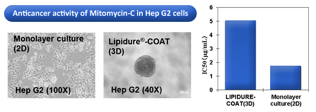
- HepG2 and other cancer cell lines
Applications
Choose your well shape for Lipidure®-Coat
Overall morphology of organoids in a culture can be altered significantly by whether cells are reaggregated in a V-bottom or U-bottom well (see Mellough et al 2019).

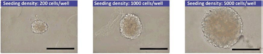
Superior Spheroid Formation
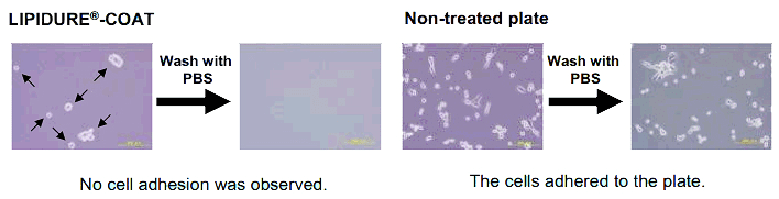
A single spheroid is generated in the Lipidure®-COAT U-bottom well, while numerous satellite spheroids are found in the competitor’s plates. This phenomenon shows that a lot of cells adhered to the competitor’s “low-adhesion” surface.

| Seeding Number (cells/well) | Diameter of Spheroid (μm) | |
|---|---|---|
| Lipidure-COAT | Competitor | |
| 1,000 | 185 ± 12 | 161 ± 9 |
| 10,000 | 423 ± 7 | 323 ± 46 |
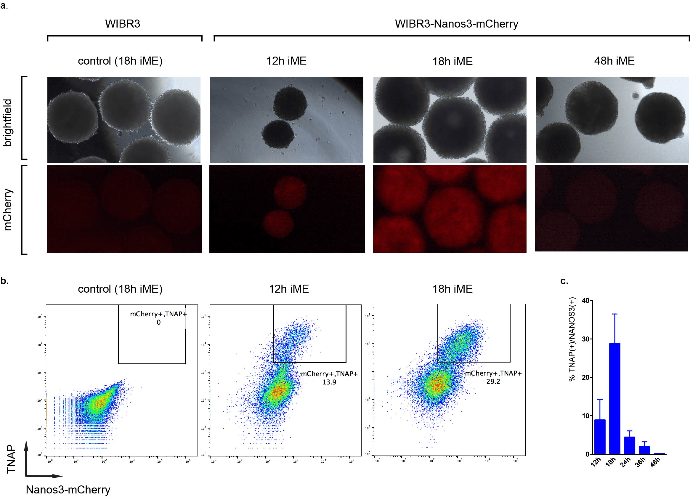
"We noticed a significantly reduced number of satellite spheroids and increased efficiency of differentiation with Lipidure U-bottom plates compared to other brands for the induction of primordial germ cell-like cells (PGCLCs)"
Organoid Culture

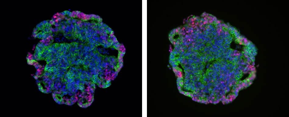
Images courtesy of Dr. Valeria Chichagova (Newcells Biotech) and Prof. Lako (Newcastle University).
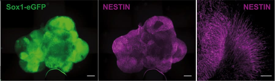
Retinal organoids continuously grown on Lipidure-coated plates from the start of differentiation. Immunofluorescence images of 150 day old organoids show rod and cone photoreceptors containing outer segments on the apical side and synaptic terminals at the base. Muller glial cells span across the entire thickness of retinal neuroepithelium.
“The Lipidure-coated plates provided by AMSBIO were extremely useful for generating with ease large numbers of homogeneous retinal organoids which responded to light and contained all the key retinal cell types.”
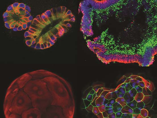
Day 2: Uniform spherical aggregates

Day 4: Ruffling, indicating formation of vesicles

Fig. Aggregation and early organoid development of inner ear organoids on U-bottom Lipidure®-Coat Wells – courtesy of the Hashino lab, Indiana University School of Medicine

Related Products & Services

Featured Citations
Self-organizing optic-cup morphogenesis in three-dimensional culture.
Eiraku, M. et al. Nature, 2011, 472 (7341), 51. Highly cited! (over 1 thousand times) - U bottom plates
Human‐induced pluripotent stem cells generate light responsive retinal organoids with variable and nutrient‐dependent efficiency.
Hallam, D. et al. Stem Cells, 2018, 36 (10), 1535-1551. U bottom plates
Systematic Comparison of Retinal Organoid Differentiation from Human Pluripotent Stem Cells Reveals Stage Specific, Cell Line, and Methodological Differences.
Mellough, C. B., Collin, J., Queen, R., Hilgen, G., Dorgau, B., Zerti, D., ... & Lako, M. (2019). Stem cells translational medicine. U and V bottom plates
A biodegradable scaffold enhances differentiation of embryonic stem cells into a thick sheet of retinal cells.
Singh, D. et al. Biomaterials, 2018, 154, 158-168. U bottom plates
Retinal organoids from pluripotent stem cells efficiently recapitulate retinogenesis.
Völkner, M. et al. Stem Cell Reports, 2016, 6 (4), 525-538. U bottom plates
iPSC-derived human microglia-like cells to study neurological diseases.
Abud, E. M. et al. Neuron, 2017, 94 (2), 278-293. V bottom plates
An organoid-based model of cortical development identifies non-cell-autonomous defects in Wnt signaling contributing to Miller-Dieker syndrome.
Iefremova, V. et al. Cell reports, 2017, 19 (1), 50-59. U bottom plates
The ubiquitin ligase LIN41/TRIM71 targets p53 to antagonize cell death and differentiation pathways during stem cell differentiation.
Nguyen, D. T. T. et al.Cell death and differentiation, 2017, 24 (6), 1063. U bottom plates
The Ca2+/Mn2+-transporting SPCA2 pump is regulated by oxygen and cell density in colon cancer cells.
Jenkins, J. et al.Biochemical Journal, 2016, 473 (16), 2507-2518. U bottom plates
Investigation of brain tissue infiltration by medulloblastoma cells in an ex vivo model.
Neve, A. et al. Scientific reports, 2017, 7 (1), 5297. U bottom plates
Antineoplastic effect of 1α, 25 (OH) 2D3 in spheroids from endothelial cells transformed by Kaposi’s sarcoma-associated herpesvirus G protein coupled receptor.
Suares, A. et al. The Journal of steroid biochemistry and molecular biology, 2019, 186, 122-129. Flat bottom plates
Foxd3 promotes exit from naive pluripotency through enhancer decommissioning and inhibits germline specification.
Respuela, P. et al.Cell stem cell, 2016, 18 (1), 118-133. U bottom plates