Wnt Recombinant Proteins & Reporter Stable Cell Lines
Cell Signalling and Organoid Culture (Wnt3a)
Wnt are a family of cysteine-rich secreted polypeptides (more than 16 mammalian family members) involved in several important cell functions such as cell-cell communication, proliferation, migration, polarity, survival and self-renewal.
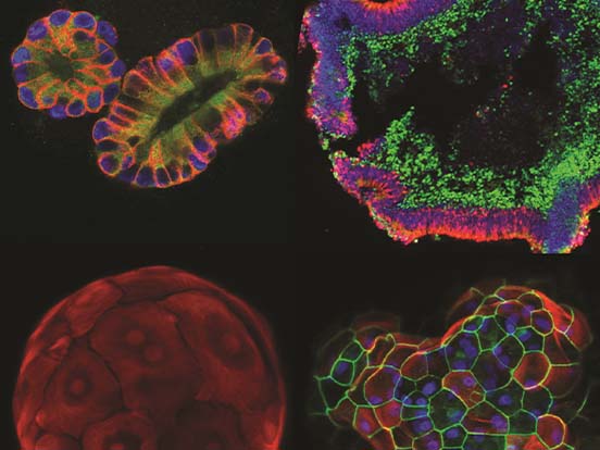
Wnt signals, in particular Wnt3a, play an important role in the ability of organoids to expand. Wnt3a is an essential niche component for maintaining the proliferation of Lgr5+ stem cells in various organoids such as the small intestine, large intestine, stomach, pancreas and liver.
Additionally, loss of activation of Wnt expression is associated with alteration of cell fate, morphogenesis and mitogenesis. Hence, diversity in functions of Wnt signalling overlaps between field of developmental biology and field of oncology leading to interdisciplinary research.
Highly Stable Recombinant Wnt3a for Organoid Culture
Our recombinant mouse and human Wnt3a have been successfully used for culture of Lgr5+ organoids. These proteins are expressed in mammalian systems, as bacterial systems can’t add polysaccharide or lipid chains to the Wnt proteins. These chains are essential for Wnt protein to be functional, so Wnt proteins can ONLY can be functional when expressed from mammalian cells.
We now supply our recombinant Wnt proteins with a new Wnt stabilizer that can significantly extend the activity of this protein, extending half-life of Wnt3a in serum-free medium to about 24-30 hours (in contrast to half-life of around 2 hours without stabilizer). Working concentration for our mouse recombinant Wnt3a (75% purity) is 30-100 ng/mL for organoid formation.
Canonical Wnt Signaling Pathway
Among the most studied is a canonical Wnt signaling pathway that has been remained well conserved among diverse species throughout the process of evolution. The key restriction point in canonical Wnt signalling is initiated by Wnt binding to Frizzled receptors.
Frizzled receptors are composed of seven transmembrane regions, extracellular Wnt binding domain and intracellular C-terminal tail. There exist eight mammalian frizzled receptors that are capable of binding multiple different Wnts. Similarly, multiple different Wnts are able to bind several different Frizzled receptors. This complexity of ligand-receptor binding is even further increased by co-receptors such as proteoglycans, particularly heparin-like glycosaminoglycans. The importance of glycosaminoglycans was highlighted by finding that embryonic injection of heparinase results in patterning defects. Therefore, loss of heparin-like glycosaminoglycans may results in Wnt proteins to be atypically diffused and have concentration in which they are no longer effective
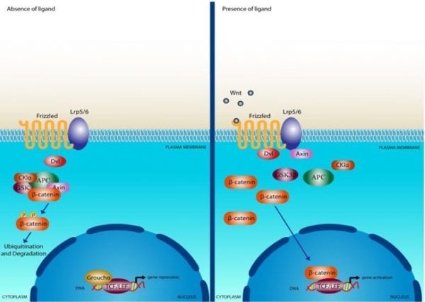
The signal from Wnt binding to frizzled receptor is further passed on to the recruited signaling molecule – Dishevelled (Dvl). Dvl proteins are present in the cytoplasm and contain protein-protein interaction domain that allows them to interact with other signaling molecules such as glycogene synthase kinase-3 (GSK-3) a cytoplasmic serine-threonine kinase involved in glycogen metabolism. The interaction of Dvl with GSK3 leads to an inhibition of GSK3 activity that in turn averts the turnover of β-catenin causing β-catenin accumulation.
β-catenin is a 90 kDa protein that contains at the N-terminus phosphorylation sites and binding site for α-catenin. In the middle are present 13 repeats creating together one superhelix that acts as a positively charged groove. This groove then in turn interacts with APC and TCF transcription factors as well as with cadherins. Complexes of β-catenin with TCF and cadherins were shown to be located in the nucleus while complexes of β-catenin with APC were found in the cytosol.
APC is a 300 kDa protein with several different functions and together with Wnt signaling is especially important in the cancer research field particularly in sporadic or inherited colon cancers. Mainly because APC is able to regulate levels of β-catenin and loss of APC function leads to a positive Wnt signaling.
Lastly, once the β-catenin forms complex with TCF or with lymphocyte enhancer binding factor 1 (LEF-1) transcription factor, it then targets responsive genes involved in migration or adhesion for example Myc, Cyclin D1, TCF-1, MMP-7, Axin-2, CD44, PPAR-δ and others.
Wnt Recombinant Proteins
AMSBIO offers a range of Wnt human and mouse recombinant proteins in high (85 – 90%) and low (75%) purity. These human recombinant proteins are purified from HEK293 cells while the mouse proteins are expressed in CHO cells. Both are suitable for various cell based assays and treatments
Your Wnt3a in the TOP/FOP Flash Reporter Assay showed higher activity
at the same concentration than the industry leader
| Description | Purified From | Purity | Application | Pack Size | Catalogue No. |
|---|---|---|---|---|---|
| Mouse Recombinant Wnt3a | CHO | 75% | Functional studies, WB, ELISA | 2 µg | AMS.rmW3aL-002 |
| 10 µg | AMS.rmW3aL-010 | ||||
| 85-90% | 2 µg | AMS.rmW3aH-002 | |||
| 10 µg | AMS.rmW3aH-010 | ||||
| Human Recombinant Wnt3a | Human | 75% | Functional studies, WB, ELISA | 2 µg | AMS.rhW3aL-002 |
| 10 µg | AMS.rhW3aL-010 | ||||
| 85-90% | 2 µg | AMS.rhW3aH-002 | |||
| 10 µg | AMS.rhW3aH-010 | ||||
| Human/Mouse Recombinant Wnt5a | CHO | 75% | Functional studies, WB, ELISA | 2 µg | AMS.rhmW5aL-002 |
| 10 µg | AMS.rhmW5aL-010 | ||||
| 85-90% | 2 µg | AMS.rhmW5aH-002 | |||
| 10 µg | AMS.rhmW5aH-010 | ||||
| Mouse Recombinant Wnt3a with stabilizer | CHO | 75% | Functional studies, WB, ELISA | 2 µg | AMS.rmW3aL-002-stab |
| 10 µg | AMS.rmW3aL-010-stab | ||||
| 85-90% | 2 µg | AMS.rmW3aH-002-stab | |||
| 10 µg | AMS.rmW3aH-010-stab | ||||
| Human Recombinant Wnt3a with stabilizer | Human | 75% | Functional studies, WB, ELISA | 2 µg | AMS.rhW3aL-002-stab |
| 10 µg | AMS.rhW3aL-010-stab | ||||
| 85-90% | 2 µg | AMS.rhW3aH-002-stab | |||
| 10 µg | AMS.rhW3aH-010-stab | ||||
| Human/Mouse Recombinant Wnt5a with stabilizer | CHO | 75% | Functional studies, WB, ELISA | 2 µg | AMS.rhmW5aL-002-stab |
| 10 µg | AMS.rhmW5aL-010-stab | ||||
| 85-90% | 2 µg | AMS.rhmW5aH-002-stab | |||
| 10 µg | AMS.rhmW5aH-010-stab |
Wnt Reporter Stable Cell Lines
AMSBIO also has a range of Wnt reporter stable cell lines. Customers can currently choose between HEK293, HCT-116, SW480 or NIH3T3 cell lines. All have available matching controls and can be ordered also as a package in pairs.
Applications
- Evaluate Wnt protein bioactivity
- Screen anti-Wnt compounds/antibodies
- Screen Wnt signalling enhancer
- Evaluate Wnt protein stabilizer
| Description | Feature | Catalogue No. |
|---|---|---|
| HEK293 Wnt reporter stable cell line (Mutant - Control) | This cell line expresses firefly luciferase in background but won’t have response to Wnt stimulation. Constantly express GFP as control of cell numbers | AMS.WRHEK293M |
| HEK293 Wnt reporter stable cell line (Active) | This Wnt reporter cell line is designed to monitor the activity of Beta-catenin based Wnt signal transduction pathway. | AMS.WRHEK293A |
| Paired HEK293 Wnt reporter stable cell lines (Control + Active) | Combination of two cell lines (Control AMS.WRHEK293M + Active AMS.WRCL293A). The control cell line is designed to show background luciferase activity for 293 Wnt TCF Reporter Cell Line-Active. The active Wnt reporter cell line is designed to monitor the activity of Beta-catenin-based Wnt signal transduction pathway. | AMS.WRHEK293P |
| HCT-116 Wnt reporter stable cell line (Control) | This Wnt reporter cell line is a control cell line, designed to show background luciferase activity for 116 Wnt TCF Reporter Cell Line-Active. | AMS.WRHCT116M |
| HCT-116 Wnt reporter stable cell line (Active) | Wnt reporter cell line is designed to monitor the activity of Beta-catenin-based Wnt signal transduction pathway. | AMS.WRHCT116A |
| Paired HCT-116 Wnt reporter stable cell lines (Control + Active) | Combination of two cell lines (Control AMS.WRHCT116M + Active AMS.WRCL116A). The control cell line is designed to show background luciferase activity for Wnt TCF Reporter Cell Line-Active. The active Wnt reporter cell line is designed to monitor the activity of Beta-catenin-based Wnt signal transduction pathway. | AMS.WRHCT116P |
| SW480 Wnt reporter stable cell Line (Mutant - Control) | Wnt reporter cell line is designed to monitor the activity of Beta-catenin-based Wnt signal transduction pathway. This human colorectal carcinoma cell line hosts CMV promoter, a mutant TCF transcriptional response element, luciferase gene, and GFP gene. | AMS.WRSW480M |
| SW480 Wnt reporter stable cell line (Active) | Wnt reporter cell line is designed to monitor the activity of Beta-catenin-based Wnt signal transduction pathway. This human colorectal carcinoma cell line hosts the TCF transcriptional response element, luciferase gene, and GFP gene. | AMS.WRSW480A |
| Paired SW480 Wnt reporter stable cell lines (Control + Active) | Combination of two cell lines (Control AMS.WRSW480M + Active AMS.WRSW480A). The control cell line is designed to show background luciferase activity for Wnt TCF Reporter Cell Line-Active. | AMS.WRSW480P |
| NIH3T3 Wnt reporter stable cell line (Mutant -Control) | This cell line expresses firefly luciferase in background but won’t have response to Wnt stimulation. Constantly express GFP as control of cell numbers | AMS.WRNIH3T3M |
| NIH3T3 Wnt reporter stable cell Line (Active) | This Wnt reporter cell line is designed to monitor the activity of Beta-catenin based Wnt signal transduction pathway. | AMS.WRNIH3T3A |
| Paired NIH3T3 Wnt reporter stable cell lines (Control + Active) | The package contains two cell lines (Control AMS. WRNIH3T3A + Active AMS. WRNIH3T3M). The control cell line expresses firefly luciferase in background but won't respond to Wnt stimulation. The active cell line expresses firefly luciferase in response to Wnt stimulation. Constantly express GFP as control of cell numbers. | AMS.WRNIH3T3P |
Wnt Antibodies
AMSBIO offers a range of polyclonal Wnt antibodies produced in-vivo, suitable for ELISA, IHC and Lateral Flow applications. In addition to antibodies AMSBIO also offers human and mouse ELISAs used for detection of several different Wnt proteins.
| Description | Source | Reactivity | Isotype | Application | Pack Size | Catalogue No. |
|---|---|---|---|---|---|---|
| Wnt-1 Rabbit Polyclonal antibody | Rabit | Human | Rabbit IgG | ELISA, Lateral Flow, IHC, WB | 100 μg | TA319125 |
| Wnt-3a Rabbit Polyclonal antibody | Rabit | Human | Rabbit IgG | ELISA, Lateral Flow, IHC, WB | 100 μg | TA328475 |
| Wnt-5 Rabbit Polyclonal antibody | Rabit | Human, Mouse, Rat, Bovine, Primate | Rabbit IgG | ELISA, Lateral Flow, IHC, WB | 100 μg | TA337059 |
| Wnt-6 Rabbit Polyclonal antibody | Rabit | Human, Mouse, Rat | Rabbit IgG | ELISA, Lateral Flow, IHC, WB | 100 μg | TA318924 |
| Wnt-8b Rabbit Polyclonal antibody | Rabit | Human, Primate, Bat, Rat, Dog, Horse | Rabbit IgG | ELISA, Lateral Flow, IHC, WB | 50 μg | TA317525 |
Selected Wnt ELISA Kits
Wnt ELISA kits allow for the in vitro quantitative determination of various mouse and human Wnt proteins concentrations in serum, plasma, urine, tissue homogenates, cell culture supernatants and other biological fluids. These immunoassay kits are only for research use and not for therapeutic or diagnostic applications.
| Description | Reactivity | Pack Size | Catalogue No. |
|---|---|---|---|
| Wnt-1 Human ELISA | Human | 96 Test/kit | AMS.E2985h |
| Wnt-1 Mouse ELISA | Mouse | 96 Test/kit | AMS.E2985m |
| Wnt-3a Human ELISA | Human | 96 Test/kit | AMS.E2182h |
| Wnt-3a Mouse ELISA | Mouse | 96 Test/kit | AMS.E2182m |
| Wnt-5a Human ELISA | Human | 96 Test/kit | AMS.EH1164 |
| Wnt-5a Mouse ELISA | Mouse | 96 Test/kit | AMS.EM0497 |
| Wnt-5b Human ELISA | Human | 96 Test/kit | AMS.E1558h |
| Wnt-5b Mouse ELISA | Mouse | 96 Test/kit | AMS.E1558m |