General Questions
What is 3D Cell culture?
What are the benefits of electrospun scaffolds?
What is the Mimetix scaffold?
The Mimetix scaffold consists of highly uniform electrospun fibres that are fabricated into non-woven, biomimetic scaffolds to support the three-dimensional growth of cells. It is made of the medical-grade polymer poly-L-lactide (PLLA) and can therefore be used to facilitate translation of research findings into clinical use. The Mimetix scaffold is provided in formats that are easy to use and compatible with standard equipment and processes; with minimal protocol adaption required to switch from 2D to 3D.
What distinguishes the Mimetix scaffold from other products on the market?
The Mimetix scaffold provides a true 3D environment � rather than a roughened 2D surface � in easy-to-use formats. It guarantees reproducible fibre diameters at standard deviations of <10% well-to-well and batch-to-batch; minimising experimental variability. It has been validated with a number of different cell types including primary cells and stem cells. The Mimetix scaffold comes in ready-to-use, sterile, standard-size plates, which are compatible with standard laboratory liquid handling robotics and automated image analysis equipment, such as plate readers.
What if I need something different?
We can work with you to develop scaffolds to suit your specific application requirements. We have experience with a range of synthetic polymers with different degradation properties and are able to tune the fibre diameter to create scaffolds with pore sizes to suit different cell types. Our other capabilities include incorporation of bioactive molecules into the fibres and coating of any of our products with a choice of 29 different peptide motifs to promote cell attachment, growth and differentiation.
Which cell types has the Mimetix scaffold been tested with?
The Mimetix scaffold has been tested with a variety of human primary cells, human cancer cell lines, and human stem cells. The table below lists the cell types which have been successfully cultured in Mimetix to date.
Sterilisation
Is the Mimetix scaffold supplied sterile?
Yes, the Mimetix scaffolds are supplied gamma-irradiated in individually-sealed plastic wrapping. The scaffolds are sterile as long as the plastic wrapping remains intact.
Can you autoclave the Mimetix scaffold?
No, the Mimetix scaffold cannot be autoclaved due to the low glass transition temperature of poly-L-lactide (60-65 �C).
Can you sterilise the Mimetix scaffold with 70% ethanol?
Sterilisation with ethanol is not recommended for formats which allow for scaffold removal (6- and 12-well), as ethanol at higher concentrations (50-70%) causes the scaffolds disks to shrink (by about 10% in 70% ethanol and 5% in 40% ethanol). Shrinkage has not been observed for the 96-well format, for which the scaffold is laser-welded into the well plate.
Usage and Handling
How do you prepare the Mimetix scaffold for cell seeding?
The Mimetix scaffold needs to be wetted with ethanol in order to allow a cell suspension to access the pores. For best results, add 1 mL 20% ethanol to each well of a 12-well plate or 200 �L to each well of a 96-well plate, and allow the ethanol to soak into the membrane. This should wet the scaffolds evenly. Next, aspirate ethanol carefully from the edge of the insert, being careful not to touch the membrane, wash the scaffold 1-2x with serum-containing cell culture medium, and add the cell suspension. We recommend seeding 50,000 to 100,000 cells suspended in 1 mL into a 12-well plate and 10,000 to 20,000 cells suspended in 200 �L into a 96-well plate as a general guideline; or alternatively, whichever cell number the experiment to be conducted requires. For long-term experiments, the medium should be exchanged every 2-3 days. If you receive the scaffold as loose discs, make sure you remove them from their backing paper.
Can the scaffolds be used in serum-free medium?
Serum contains components which help cells to attach to surfaces. The absence of serum may therefore hinder cell attachment to our scaffolds for certain cell types. If you do experience problems with culturing cells in our scaffolds using serum-free medium, consider the following options: 1) It is sometimes possible to condition cells over time to grow in lower concentrations of serum. 2) Consider using a medium with serum supplements (for more information click here). 3) Various coatings can facilitate cell adhesion in low serum conditions, such as poly-L-lysine. Please refer to the last question in this section for more information.
How do you estimate cell confluency within the Mimetix scaffold?
Confluency and cell viability can be estimated with optical or fluorescence microscopy using appropriate dyes for cell staining (e.g. neutral red). Note that some hydrophobic dyes may adsorb onto the scaffold. Quantitative, colorimetric assays (see next section) are also compatible and can provide a more accurate measurement of the cell number within a scaffold.
How long can you culture cells within the Mimetix scaffold?
Depending on the cell type and the method of analysis to be performed, the Mimetix scaffold in our well plate-formats is recommended for cell culture experiments lasting up to 10 days. For longer experiments, a hanging-insert format is available, allowing cells to be cultured for up to 4 weeks.
Can you retrieve cells from the Mimetix scaffold?
Yes, cells can be removed from the Mimetix scaffold, optimal cell recovery is obtained following these steps:
- Wash the cells twice with PBS
- Add 40 �l 1xTrypsin per well
- Incubate the plate at 37�C for 6 minutes
- Shake the plate vigorously for 2 minutes on a plate shaker
- Harvest the cells
Does growing cells on the Mimetix scaffold trigger any cellular processes?
Yes, it is well known that cells grown in 3D demonstrate different behaviours to those grown in 2D*. It is possible that the architecture of the scaffold could promote certain pathways over others, and that these differences are representative of in vivo situations than 2D. For example, recent data using a human breast cancer cell lines (HMT3909S8) in our Mimetix scaffold have shown differences in proliferation rate and response to staurosporine-induced apoptosis. We are always interested in receiving feedback on the performance of our scaffolds with different cell types and are in the process of setting up a database to be able to answer this question in more detail in the future.
*Rimann et al., Curr. Opin. Biotechol. 2012, 23, 803-809.
Can you coat the Mimetix scaffold?
Yes, the Mimetix scaffold can be coated with most conventional methods. Depending on the coating agent used, this might slightly increase fibre diameters and decrease pore size.
Format and Properties
What formats are available?
Our randomly-oriented Mimetix scaffold is available with fibres of 2 �m and 4 �m diameter (correlating with pore sizes of 10-18 �m and 15-31 �m, respectively) and in two different thicknesses; 50 �m and 100 �m. It can be supplied in a standard 12-well format, a 6-well format with hanging inserts, and 96-well format. Alternatively, the scaffold can be shipped as a sheet (220 mm x 350 mm) for customised cutting, or as pre-cut disks with diameters between 2 and 50 mm. Our aligned Mimetix scaffold is available with a fibre diameter of 2 �m, mounted on 12-well crown inserts. All formats include the plate and lids.
Which scaffold format should I use?
The best scaffold format to use depends on a variety of factors, such as cell type, duration of culture, desired penetration depth into the scaffold, and assays to be performed. Our 12-well format is suitable for short-term cell cultures and confocal microscopy, as it allows for removal of the scaffold. Hanging inserts are suited for longer culturing periods or cell migration experiments; with cells in the scaffolds being exposed to medium from above and below. High-throughput experiments can be performed with our 96-well plate format. In this case, however, the scaffolds cannot be removed from the wells.
What is the Mimetix scaffold made of?
The Mimetix scaffold is made of the FDA-approved, medical-grade polymer poly-L-lactide (PLLA).
Is the Mimetix scaffold biodegradable?
Yes, electrospun fibres made from PLLA are susceptible to hydrolytic degradation both in vitro and in vivo; however, the rate of breakdown is very slow. When used for in vitro applications the material can be considered as non-biodegradable. The rate of breakdown in vivo depends on several factors, such as the surrounding physiological environment, degree of vascularisation, mechanical stress, and the fibre diameter/scaffold thickness.
A study by Blackwood et al.* analysed the biodegradability of larger fibres and found that PLLA fibres with an average diameter of 2.1 �m remained largely intact after one year of subcutaneous implantation in rats, with some slight reduction of the fibre diameter within this period.
If you require scaffolds that degrade within a shorter time-frame or scaffolds made of non-biodegradable polymers, please contact us to discuss our bespoke service.
*Blackwood et al., Biomaterials 2008, 29, 3091�3104.
What is the pore size of the Mimetix scaffold?
Pore sizes are difficult to estimate as the scaffold is non-woven and all pores are interconnected, but a measurement of surface pores indicated that pores in the 2 �m scaffolds are roughly 10-18 �m wide, whereas the majority of pores in the 4 �m scaffolds are between 15 and 31 �m in size. The overall porosity of the scaffold is 80%. If you require scaffolds with smaller or larger pores, please contact us to discuss our bespoke service.
Which fibre diameter/pore size should I use?
Can a scaffold be made from different polymers?
We provide a bespoke service to design and develop electrospun scaffolds to suit user requirements. We have a range of polymers available, for which we can adjust the fibre diameter and scaffold thickness. Furthermore, we can prepare multi-layer or mixed membranes, coaxial fibres, aligned fibres, electrospray coatings, and spin onto 3D shapes. Please contact us or click here for more information.
Can the Mimetix scaffold be made with different pore sizes or thicknesses?
Yes, we can provide bespoke products with different fibre diameters, pore sizes, and thicknesses. Please contact us if you would like to discuss the development of electrospun scaffolds for your specific application.
Can you get reproducible data with the Mimetix scaffold?
Our randomly-oriented fibre scaffolds are highly consistent with respect to fibre diameter and are suitable for culturing a range of cell types, including primary cells and stem cells. Experiments with the HMT3909S8 primary human breast cancer cell line showed a disk-to-disk variability of only 3% to 12% for cell loading experiments and 5% to 18% for a cell count after 10 days of proliferation. Further experiments with other cell lines, including primary human hepatocytes and endothelial cells, are currently under way.
Is the Mimetix scaffold chemically resistant?
Electrospun PLLA fibres are sensitive to temperatures above 60�C, acids, and organic solvents (required for some experimental protocols for RNA and protein extraction). Cells must be removed from the scaffold and lysed prior to such analysis. As a general rule, the more hydrophobic the solvent, the more likely it is to dissolve the PLLA scaffold. Please refer to the list below for more detailed information on which solvents PLLA is compatible/incompatible with. If you are unsure, please contact us for more information or simply carry out a quick test yourself by immersing a disk in the solvent to-be-used for 1 hour and see if it disintegrates.
Incompatible solvents:
Benzene, chloroform, dichloroacetic acid, dichloromethane (DCM), dimethylformamide (DMF), isoamylalcohol, N-methyl pyrrolidone (NMP), toluene, trichloromethane.
Solvents which cause swelling:
Acetone, dimethylsulfoxide (DMSO), methylethylketone (MEK), tetrahydrofuran (THF), ethylacetate, xylene (often used in paraffin wax sectioning).
Compatible solvents:
Water, alcohols (e.g. methanol, ethanol, isopropanol), unsubstituted hydrocarbons.
*S�derg�rd et al., Prog. Pol. Sci. 2002, 1123�1136, Polymer Data Handbook, Oxford University Press, 1999.
Is the Mimetix scaffold appropriate for use in bioreactors?
The Mimetix scaffold can be mounted onto retaining rings for use in the Quasi-Vivo� bioreactor available from Kirkstall Ltd. Please contact us if you would like to discuss the development of the Mimetix scaffold for use in other bioreactor formats.
Is the Mimetix scaffold reuseable?
No, the Mimetix scaffolds are disposable and designed for single-use.
Analysis
Which analysis techniques can be used for the Mimetix scaffold?
The Mimetix scaffold is compatible with a variety of analysis techniques, including cell-based assays (please refer to the questions at the end of this section for more information), fluorescence microscopy, optical microscopy (to some extent), RNA extraction and real-time qPCR, protein extraction and Western blotting, as well as histological techniques (e.g. H&E staining) which do not involve the use of solvents. Please contact us if you are unsure whether the Mimetix scaffold would work for the technique that you are planning to use.
Can you see cells growing within the Mimetix scaffold with an optical microscope?
Observing cells within Mimetix using a traditional bench-top optical microscope can be challenging because of the semi-transparent nature of the scaffold. We recommend the use of a fluorescent microscope for imaging.
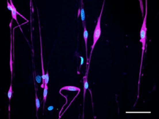
How do you image cells growing in vitro within the Mimetix scaffold?
The ideal tool to visualize cells growing in 3D in the Mimetix scaffold is confocal microscopy (see here for some examples of 3D imaging). The Mimetix scaffold is not autofluorescent at standard excitation/emission wavelengths. Cellular components can be stained using standard protocols (please refer to our protocols section), for example nuclei with DAPI or TO-PRO3, or actin with Alexa Fluor� phalloidin. The Mimetix 96-well plate furthermore has a base of superior optical clarity, which was especially designed for high-content imaging systems. The use of mounting media, for example VECTASHIELD�, can help to increase the translucency of the scaffold
How can I determine the number of cells growing in a scaffold and generate a growth curve?
Cells numbers in 3D cultures can be assessed by quantitative cell viability assays, which usually measure DNA content, ATP or metabolic activity. Assays which have been successfully used for the Mimetix scaffold are CellTiter-Glo� from Promega (measuring ATP), Alamar Blue assays, and MTS assays (both measuring metabolic activity; available from a number of suppliers). While the first two assays are based on a luminescent/fluorescent read-out and can be analysed directly in the Mimetix plate, the MTS assay is an absorbance-based assay for which the scaffolds give rise to a background signal. In this case, well contents need to be transferred to a new 96-well plate prior to read-out.
Does the Mimetix scaffold give rise to autofluorescence?
No, no significant levels of autofluorescence are observed for the Mimetix scaffold at standard excitation wavelengths.
Will absorbance-based assays work with Mimetix?
The Mimetix scaffolds gives rise to a background signal in absorbance-based assays. We hence recommend the use of fluorescence- or luminescence-based assays. Alternatively, well contents can be transferred to a new 96-well plate for an absorbance-based read-out.
Will fluorescence- and luminescence-based assays work with Mimetix?
Yes, fluorescence- and luminescence-based assays are compatible with Mimetix. No significant levels of autofluorescence are observed at standard excitation wavelengths.